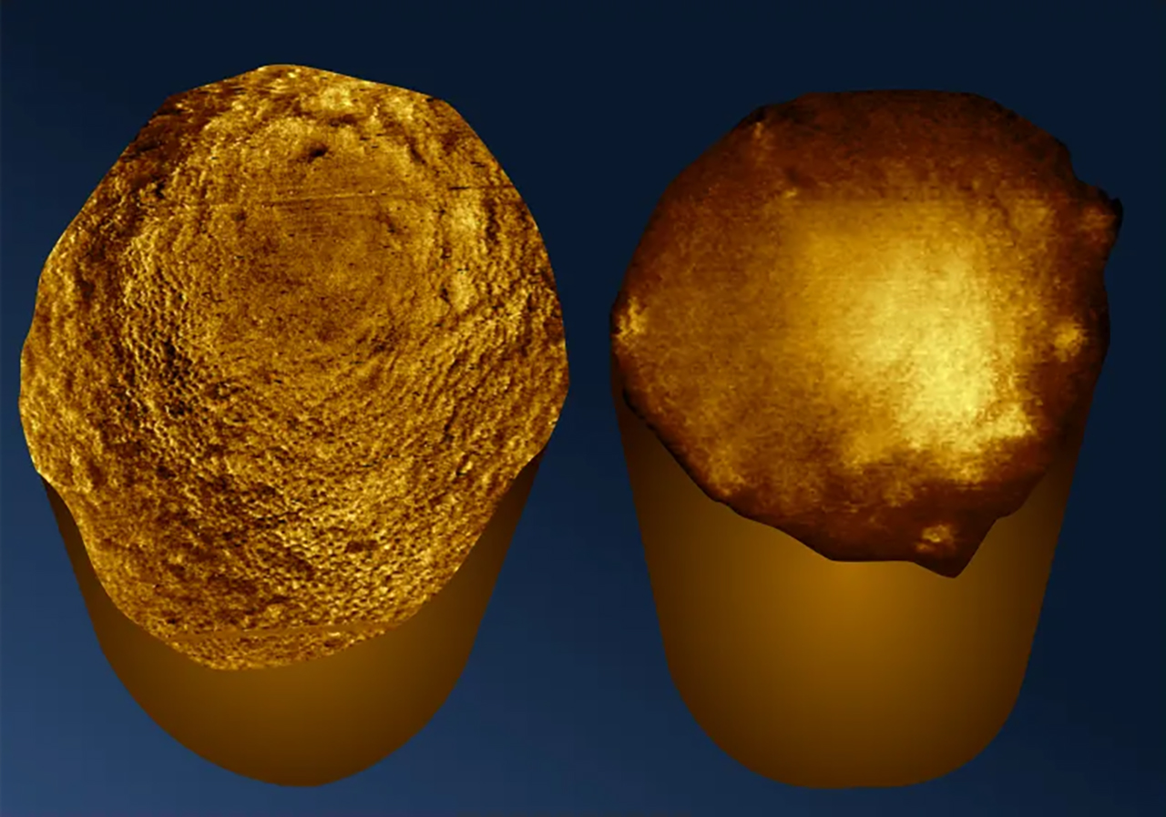
Scientists have captured detailed images of the poles at the ends of rod-shaped bacteria for the first time, using a method that keeps the microbes still and alive during imaging.
The process, described in a new study published in Nature Communications, uses glutaraldehyde to attach E. coli bacteria to glass. The substance binds to proteins on the bacterial surface while preserving the cells’ structure and keeping them alive.
Researchers then scanned the bacteria with atomic force microscopy, in which a probe a few nanometers wide taps along a cell to create an image. The technique is compared to a human finger reading braille.
The process took patience: the glutaraldehyde needed 20 minutes to take effect, and researchers waited 50 minutes to find some of the trapped bacteria standing upright. Getting bacteria to stand up for imaging has proved difficult across many types of experiments.
Study author Bart Hoogenboom said in a statement: “Imaging the poles of bacteria is like opening a view of the back of the moon,” adding, “We’re looking at something that no one else has seen before. The poles are where cells divide, and so, (they) contain critical information about this process. They may also be a particular target for antibiotics, with some bacteria-killing treatments preferring to attack this part of the cell.”
The study says images of the poles could help researchers understand parts of bacterial cells involved in growth and defense. The researchers say the imaging approach could also be adapted for longer observations, including successive cell divisions and biofilm formation.