The basal ganglia are groups of cells that sit deep in the brain and process motor, cognitive, mood and decision-making information. They are linked to neurodegenerative and psychiatric conditions from Parkinsonâs and Huntingtonâs disease to schizophrenia, bipolar disorder and addiction.
Now, researchers have published the worldâs most-detailed atlas of basal ganglia so far, comprising 25.5 million brain cells from humans, macaques and marmosets. The work is described in a collection of 26 papers published in Cell, Cell Reports, Cell Reports Methods, Cell Systems, Cell Genomics and Neuron.
The research includes understudied neurons1, provides insights about how some brain cells change with age2 and suggests which might be linked to diseases3.
The atlas, a collaboration between hundreds of scientists in nine countries, is part of the BRAIN Initiative Cell Atlas Network (BICAN) â a US$500-million project launched in 2022 by the US National Institutes of Health aimed at creating reference maps of mammalian brains.
âItâs a massive dictionary that we needed in the field,â says Ana Muñoz-Manchado, a neuroscientist at the University of Cádiz in Spain, who studies the basal ganglia in neurodegenerative disorders.
The basal ganglia atlas contains 21 million cells from the brains of six humans, three million cells from three rhesus and cynomolgus macaques, and 1.5 million cells from two marmosets4. The researchers also used data from previously published maps of the whole mouse brain5,6.
By comparing the basal ganglia between the primate species, the researchers identified 61 cell types in humans, macaques and marmosets that have been conserved over 76 million years3.
The team analysed brain tissue samples from different developmental stages across the lifespan of the species. The latest work combines information about these cells in unprecedented detail, from measurements of their gene expression and DNA modifications that control gene regulation to recordings of their electrical activity, as well as how DNA is folded inside their nuclei.
The researchers also mapped where each cell is located in the brain and designed a common framework to characterize and annotate the cells. It is âessentially a scaffold on which we can now fairly readily complete the rest of the brainâ, explains Michael Hawrylycz, a bioinformatician at the Allen Institute for Brain Science in Seattle, Washington, who took part in the project.
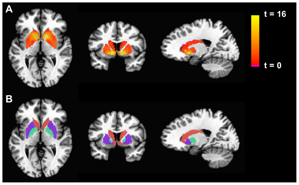
One study examined 1.1 million cells in the basal gangliaâs largest structure â known as the striatum â from 19 deceased human donors7. Unlike many other brain areas, the striatum has no neat anatomical boundaries. The researchers identified six zones in the human and macaque striatum with distinct molecular signatures, each containing distinct populations of neurons and glial cells.
âThe cells in these zones all look the same under the microscope, but have very strong transcriptional differences and are strikingly consistent from person to person,â said co-author Evan Macosko, a neurobiologist at the Broad Institute of Harvard and MIT in Cambridge, Massachusetts, in a press release from the institute.
Macosko and his colleagues also found that gene expression patterns that distinguished each zone from the other became increasingly similar with age, particularly in brain regions affected by Huntingtonâs and Parkinsonâs disease.
Another study analysed nearly four million cells in the striatum and prefrontal cortex from 178 deceased adult donors between the ages of 27 and over 902. The researchers found that the cellular make-up of the two brain regions varied between people, particularly for non-neuronal cells known as glial cells. This underscores the need for brain cell atlases to account for these individual differences.
doi: https://doi.org/10.1038/d41586-026-03565-3
Dalley, R. et al. Cell Rep. https://doi.org/10.1016/j.celrep.2026.118269 (2026).
Burger, S. C. et al. Cell 189, 7369â73856 (2026).
Johansen, N. J. et al. Cell 189, 7309â7329 (2026).
The BRAIN Initiative Cell Atlas Network (BICAN). Cell 189, 7289â7308 (2026).
Langlieb, J. et al. Nature 624, 333â342 (2023).
Yao, Z., et al. Nature 624, 317â332 (2023).
Kraft, A. W. et al. Cell 189, 7350â7368 (2026).
Record-breaking Ebola outbreak: phone data reconstruct first weeks of spread
Rise of Germanic and Celtic languages is written in ancient DNA
The grand plan to transform psychiatry â by personalizing mental-health care
New culprit in early Alzheimerâs: bloodâbrain barrier blocks protective proteins
Is social media changing childrenâs brains? Hereâs what the science says
Fruit, flies and getting frisky: how food odours trigger sexual arousal
DeepMindâs ultra-powerful AI for genome mapping wows scientists
Move over BMI: new obesity definition is better for predicting your health
New culprit in early Alzheimerâs: bloodâbrain barrier blocks protective proteins
CIB invites distinguished Strategic Scientist, Outstanding Young Investigator, and Postdoctoral Research Fellow.
Chengdu Institute of Biology, Chinese Academy of Sciences
Applicants should meet the requirements for the National Science Fund for Excellent Young Scholars (overseas), please refer to the Part IV for details
The State Key Laboratory of Quantitative Synthetic Biology
Perelman School of Medicine at the University of Pennsylvania seek candidates for an Assistant Professor position in the…