Investigators presented several studies evaluating the diagnostic performance of artificial intelligence applications in molecular pathology at the Association for Molecular Pathology 2026 Annual Meeting & Expo.
In one study focusing on blood and lymphatic malignancies, researchers developed an artificial intelligence foundation model designated ALMA 3 that analyzes DNA methylation patterns to identify and classify blood cancers.
The investigation involved collaborative work by researchers from ALMA Genomics, the University of Pennsylvania, the Massachusetts Institute of Technology, and several other academic and clinical institutions.
The model was trained using a dataset of more than 110,000 patient samples representing 81 distinct subtypes of blood and lymphatic cancers, including categories that are challenging to differentiate using standard diagnostic techniques.During independent diagnostic testing, the ALMA 3 model detected the presence of hematolymphoid tumors with 99.5 percent sensitivity and achieved a 90.9 percent positive predictive
value when assigning specific cancer subtypes.
Study authors noted that accurate subtyping is critical for guiding prognosis and therapeutic strategies.
While the study demonstrated the capacity for epigenomic AI models to complement conventional pathology workflows, the researchers reported that further clinical validation studies remain under way to confirm performance in diagnostic settings.A separate research initiative at the University of Michigan evaluated an open-source platform, named Turnkey Precision Oncology, designed to assist pathologists in
interpreting complex sequencing data and compiling clinical reports.
The platform incorporates an artificial intelligence component called TPO-Agent, which evaluates DNA and RNA sequencing results, identifies genomic alterations, and cross-references findings against established cancer genetics databases and published scientific literature to confirm that interpretation statements are directly backed by cited evidence.When evaluated across 50 genetic alterations in samples from 10
patients, approximately 95 percent of the claims generated by TPO-Agent were verified as correctly supported by cited evidence, compared to a 65 percent support rate for a conventional artificial intelligence tool equipped with comparable search resources.
Across 28 clinical reports, the broader software platform demonstrated approximately 95 percent concordance with a clinical laboratory in identifying individual alterations, with high agreement between AI-drafted reports and expert interpretations of clinically significant genetic variants.At Memorial Sloan Kettering Cancer Center and the Icahn School of Medicine at Mount Sinai, researchers evaluated whether
artificial intelligence models could triage lung adenocarcinoma specimens to determine when rapid molecular testing is necessary.
Rapid assessment for epidermal growth factor receptor gene mutations guides decisions regarding targeted therapy, but repeated testing runs the risk of depleting small biopsy specimens and using substantial laboratory resources.
The research team integrated two digital pathology models, TAPFM and EAGLE, into an evaluation workflow where six molecular pathology fellows reviewed 80 consecutive lung adenocarcinoma cases.Reviewers using the AI triage workflow determined that rapid EGFR testing could have been safely deferred in 26 of the 80 cases, representing 33 percent of the cohort, without missing any confirmed EGFR-positive tumors.
However, the authors observed that the automated approach showed lower reliability when attempting to identify cases that could forego rapid testing for certain gene fusions.
These findings indicate that while image-based triage could reduce unnecessary rapid testing and conserve diagnostic tissue, human-in-the-loop oversight and further model refinements remain necessary before implementation.Investigators at Beth Israel Deaconess Medical Center and Harvard Medical School analyzed whether foundation models could distinguish between hepatocellular carcinoma and intrahepatic
cholangiocarcinoma using standard microscope slides.
The two primary liver cancer types originate from distinct cell lineages and require different clinical interventions.
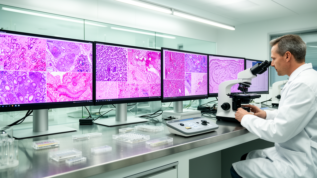
Using 50 digitized whole-slide images from The Cancer Genome Atlas public repository, the researchers evaluated a zero-shot pathology language foundation model operating without disease-specific pre-training.The computational model identified morphological patterns associated with cancer-type-specific gene expression, particularly features characteristic of normal hepatocytes.
These computational image classifications were corroborated by matching RNA sequencing data from the same tumor specimens.
The investigators reported that such models may enable the extraction of molecular and lineage-specific information directly from standard diagnostic tissue slides without requiring additional laboratory processing.Finally, a study led by researchers at Stanford University assessed the capacity of a deep-learning foundation model to predict the presence of specific genetic mutations from routine hematoxylin and eosin
slides of gastric adenocarcinoma.
Examining digitized tissue slides from 367 gastric cancer patients, the model evaluated visual morphological features to predict mutations across four cancer-related genes. The model achieved discrimination performance scores of 0.80 for…